
Image-to-Image Translation for Medical Images Using Flow Matching
Applying Conditional Flow Matching to translate MRI images into synthetic CT volumes for the Institute of Forensic Medicine at the University of Zurich.
Initial situation
Radiation therapy treatment planning requires CT images to compute dose distributions, as CT provides the electron density information needed for accurate dose calculation. In many clinical scenarios, however, MRI is the preferred imaging modality due to its superior soft tissue contrast and absence of ionising radiation. Generating synthetic CT (sCT) images directly from MRI would enable MRI-only workflows, eliminating the need for a separate CT acquisition and the associated registration between the two modalities. Two prior projects at the Institut for Data Science (I4DS) explored this task on a paired MRI–CT dataset provided by the IRM-UZH, using GAN-based architectures and diffusion models respectively.
Problem statement / Project goal
The existing approaches each come with notable limitations:
- Generative Adversarial Networks (GAN)-based models (ResViT) produced blurry outputs and failed to reconstruct fine bone structures such as teeth.
- Diffusion models (DDPM) achieved high image quality — domain experts could not distinguish generated slices from real CT — but required approximately two hours to generate a single volume due to the iterative sampling process.
- Both prior projects operated on individual 2D slices, leading to visible inter-slice inconsistencies when volumes are reassembled and viewed along other axes.
The goal of this project is to investigate whether Flow Matching (FM), a recent generative modelling framework, can match or exceed the image quality of diffusion models while substantially reducing inference time, and to explore extensions from 2D slice-based to 2.5D and 3D volume-based generation.
MRI–CT Pairs
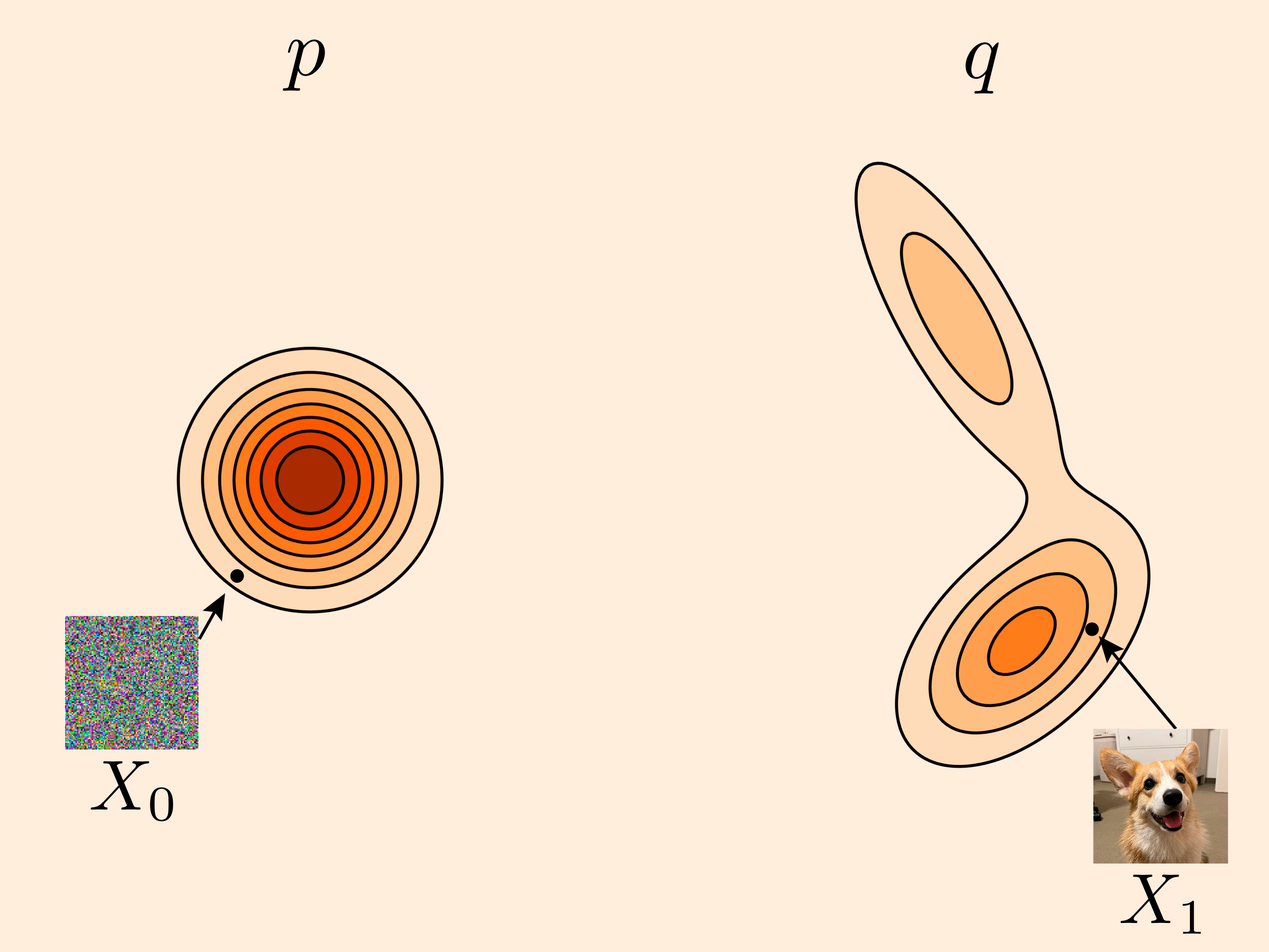
The goal is to learn a mapping from MRI images (source distribution p) to corresponding CT images (target distribution q).
Probability Path
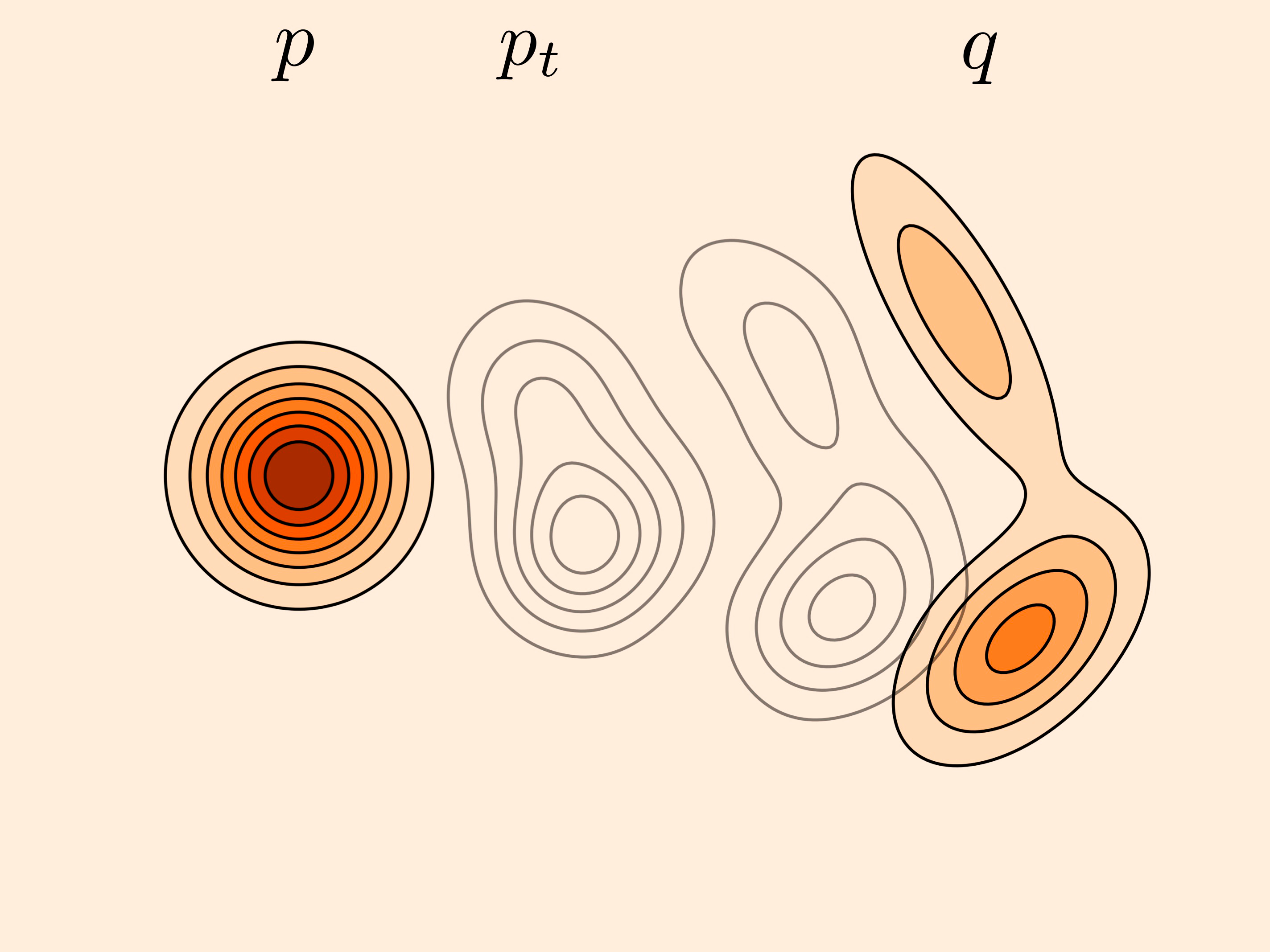
A continuous probability path p_t is constructed that smoothly interpolates between the MRI distribution at t=0 and the CT distribution at t=1.
CT Synthesis
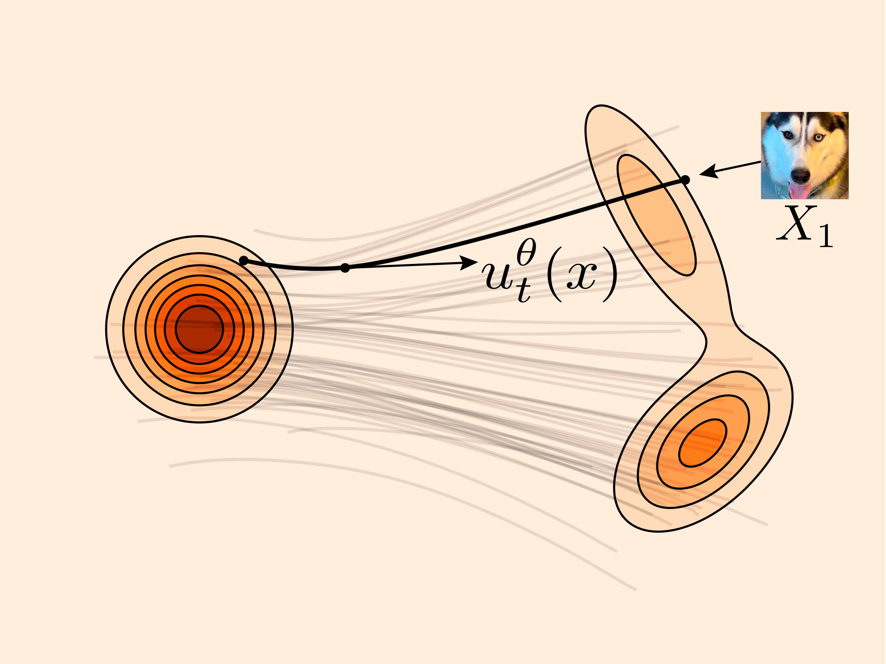
To generate a CT image, the learned velocity field is integrated from t=0 to t=1, starting from an input MRI image, producing a synthetic CT scan.
Source all images: Lipman et al., Flow Matching Guide and Code, 2024. arXiv:2412.06264
Solution developed and its benefits
This work applies Conditional Flow Matching with optimal transport paths (OT-CFM) to the MRI-to-CT translation task. A U-Net-based velocity network learns to transport samples from a Gaussian noise distribution to the CT data distribution, conditioned on the corresponding MRI input. Unlike diffusion models, FM requires only a small number of deterministic ODE integration steps at inference rather than hundreds of iterative denoising steps. The approach was systematically developed in three experimental phases — from establishing a 2D baseline through architectural exploration to 2.5D multi-slice and fully 3D volumetric models — with the aim of reducing inter-slice artefacts and improving anatomical consistency across the generated volumes.
Key terms
- keyword 1Flow Matching
- keyword 2MRI-to-CT Translation
- keyword 3Medical Image Synthesis
Customer

Institute of Forensic Medicine
Dr. Niklaus Zölch
Winterthurerstrasse 190/52
CH-8057 Zürich
any link to the company
Team
Julia Locher
Alessia Vannini
Advisor
Prof. Dr. Michael Graber
Joe Weibel