
How MRI Input Channels Affect Deep Muscle Segmentation Models
A systematic evaluation on the effectiveness of combining multiple MRI image inputs for automated muscle segmentation.
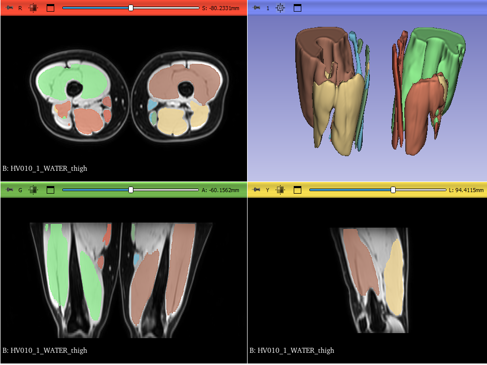
3D visualization of the resulting segmentation masks and the MRI image.
Initial situation
The quantitative analysis of muscle volume using magnetic resonance imaging (MRI) is becoming increasingly important in medical diagnostics. It provides valuable insights and plays an important role in surgical planning, rehabilitation, and injury prevention. To quantify the muscle volume, the images have to be segmented - a task that is currently mostly performed manually by trained medical professionals. As this task is time intensive and demanding, solutions to automate the segmentation process are sought after.
Problem statement
Deep-learning models offer a promising avenue for automating the segmentation process and reducing the burden on medical professionals. However, they require huge amounts of labeled data for their training. In MRI imaging, a single scan can consist of multiple output contrasts, which highlight different tissue. In theory, this would allow for more diverse input images for one single segmentation file, but historically, often only one contrast is used. A systematic evaluation of the effect of combining different contrasts as input channels could therefore be beneficial. This project aims to provide such overview by evaluating which single input channel as well as which combination of input channels yields the best segmentation result.
Study Design
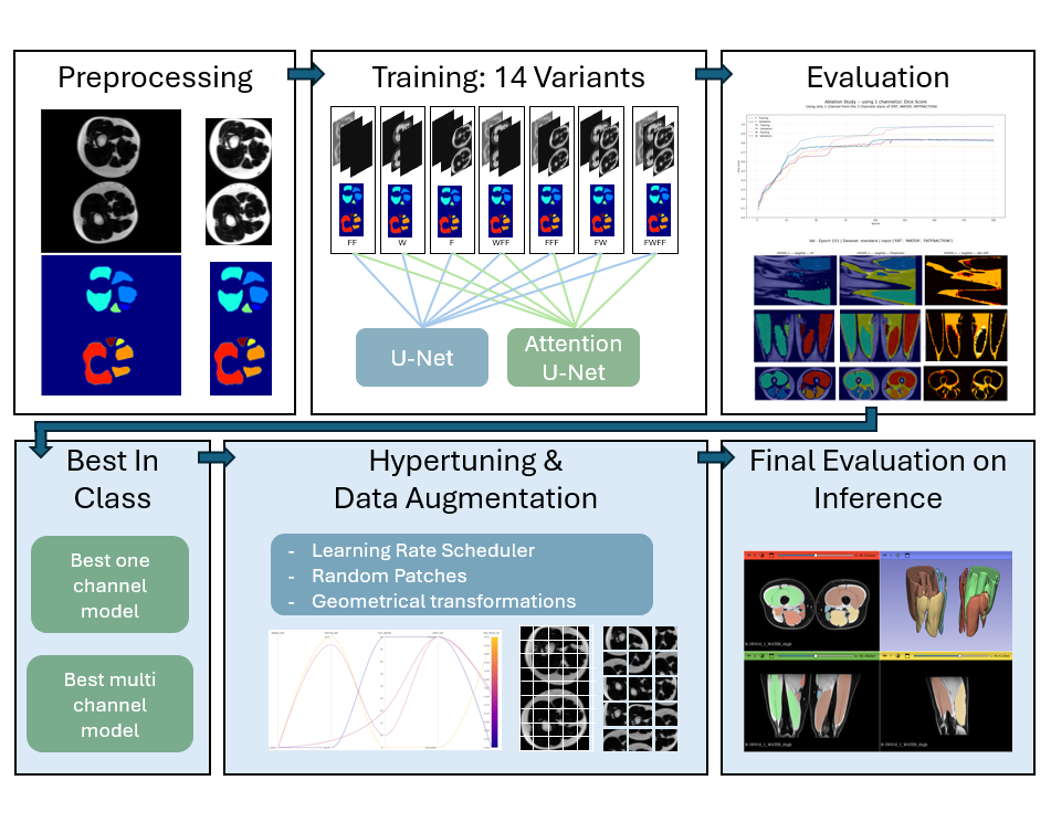
The image depicts the applied workflow: all images were preprocessed, then fed into the models and evaluated. The best two models were further improved before a final evaluation on inference data was performed to find the best model.
Training
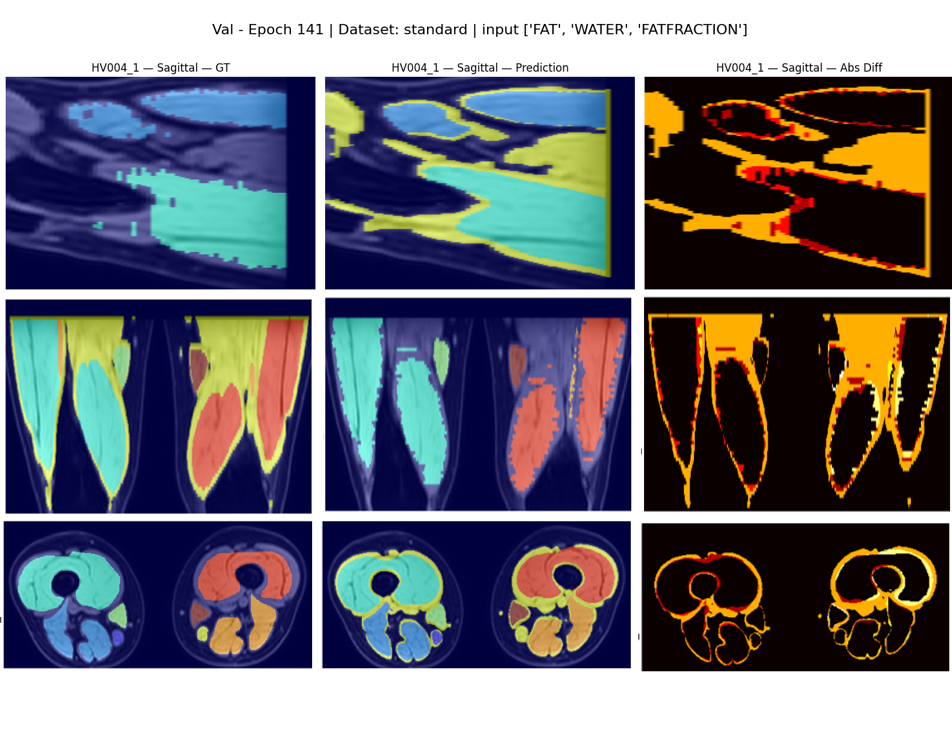
During training, the segmentations were visualized in all 3 axis every epoch to supervise the progress and make sure the model was learning correctly.
Evaluation
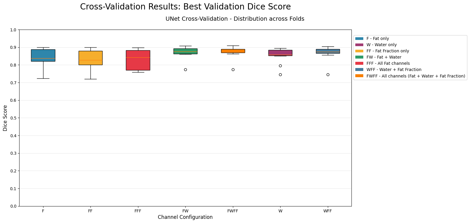
The Dice Score was used to evaluate model performance and find the best models.
Solution developed and its benefits
Two different deep learning models (U-Net and Attention U-Net) were implemented and trained with all possible combinations of one, two or three input channels. To ensure comparability between models, missing channels were replaced with an empty input channel. Out of the 7 combinations, the best one channel model and best multichannel model were further improved by adding in data augmentation and hyperparameter tuning. The final best model was then compared to a state-of-the-art automated segmentation tool, to ensure the quality of segmentation is appropriate. The chosen approach has the following benefits:
- Systematic evaluation of each type of input
- Comparable results due to the models having the same size and same amount of parameters
- Evaluation of the effect on 3D segmentation, rather than 2D
To our knowledge, this is the first study evaluating the effect of combinating different input channels for 3D segmentation, which provides a valuable stepping stone for further research in this area.
Key terms
- Language Python
- Libraries MONAI, SimpleITK, numpy
- Tools 3D Slicer, Weights and Biases
- Keywords Medical Image Analysis, Automated Segmentation, U-Net, MRI images, Dixon sequences, ablation
Customer
Balgrist Campus
Prof. Dr. Daniel Nanz
Dr. Stefan Sommer
Prof. Dr. Jörg Spörri
https://www.balgristcampus.ch/
Team
Antonia Bonfa
Alessio Lopo
Advisor
Prof. Dr. Susanne Suter